Research
Information about skin cancer and analysis patterns.
Investigación
Información sobre el cáncer de piel y el análisis de patrones.
BENIGN AND MALIGNANT TUMORS
MORPHOLOGY PATTERNS
COLOR PATTERNS
BENIGN AND MALIGNANT TUMORS
A tumor is an abnormal lump or growth of cells. When the cells in the tumor are normal, it is benign. Something just went wrong, and they overgrew and produced a lump. When the cells are abnormal and can grow uncontrollably, they are cancerous cells, and the tumor is malignant. 1,2,3
If you have been diagnosed with a malignant tumor, your doctor will create a treatment plan with you based on the stage of cancer. Early-stage cancers haven't spread much, if at all, whereas later-stage cancers have spread to more areas of the body. Determining the stage of cancer may require biopsies, surgery, and/or imaging tests. Once the cancer stage is determined, you can proceed with therapy. 2,4
If you have been diagnosed with a benign tumor, your doctor will provide reassurance that you do not have cancer. Depending on the type of benign tumor, your doctor may recommend observation or removal for cosmetic or health purposes (for instance, the tumor may be compromising an important organ in your body). 5
Characteristics of Benign Tumors
- Cells tend not to spread
- Most grow slowly
- Do not invade nearby tissue
- Do not metastasize (spread) to other parts of the body
- Tend to have clear boundaries
- Under a pathologist's microscope, shape, chromosomes, and DNA of cells appear normal
- Do not secrete hormones or other substances (an exception: pheochromocytomas of the adrenal gland)
- May not require treatment if not health-threatening
- Unlikely to recur if removed or require further treatment such as radiation or chemotherapy
Characteristics of Malignant Tumors
- Cells can spread
- Usually grow fairly rapidly
- Often invade basal membrane that surrounds nearby healthy tissue
- Can spread via bloodstream or lymphatic system, or by sending "fingers" into nearby tissue
- May recur after removal, sometimes in areas other the original site
- Cells have abnormal chromosomes and DNA characterized by large, dark nuclei; may have abnormal shape
- Can secrete substances that cause fatigue and weight loss (paraneoplastic syndrome)
- May require aggressive treatment, including surgery, radiation, chemotherapy, and immunotherapy medications
MORPHOLOGY: BEST PATTERN FOR MODELLING
Articles describe benigns by a mobile mass smooth and round with a surrounding fibrous capsule, where cells multiply slowly and cannot lead to metastasis. According to pictures, this description fits very well in nearly all of the cases, except for some rare conditions of carcinomas. 4,5
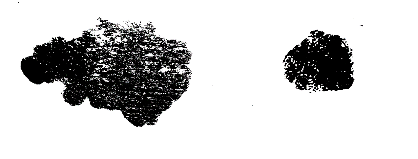
On the other side, malignant tumors have got an irregular shape with no capsule due to the fact that cells are not forming a mass, they act as individuals and they spread and divide uncontrollably, leading to metastasis. 6 Its shape is not consistent, is usually dynamic, as it can variate depending on the cancer progress and has not a defined outline due to the mentioned characteristics (Figure 1). 3,6,9,10
SUMMARY: Benign tumors have got a regular shape with a surrounding fibrous capsule that defines the location of the tumor whereas malignant tumors do have an irregular shape with no defined outline.
Figure 1. Shape difference between a malignant and a benign TUMOR. Left-photo: Malignant tumor, corresponding to a carcinoma. It’s observed an irregular shape with not a defined outline. Right-photo: Seborrhoeic Keratosis, a benign tumor with a regular shape that resembles a circle with a good defined outline.
COLOR: PLAYING AGAINST THE ODDS
Colour is not a determinant of benign or a malignant tumor as there are multiple factors that determine their color like the location, the access to blood vessels, disrupted genes, among others.
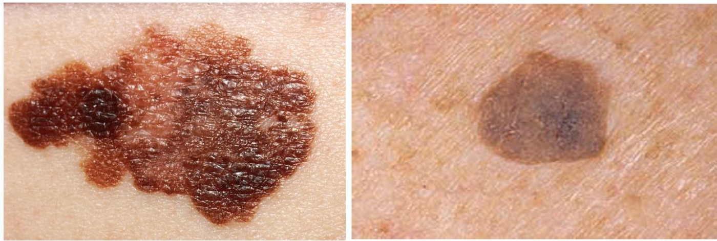
there are lots of factors to take into account and multiple variables that we cannot control. Human bias also contributes to decreasing the efficiency of looking at those patterns as it depends on the illumination of the picture, the resolution and the topology of the skin, which in fact will also affect proper classification (Figure 2)3,7,8
SUMMARY: It’s very difficult to make a recognition from color as there are multiple variables that we cannot control which affect the gradients and tonalities. Human bias also contributes to this low level efficiency.
Figure 2. Colour difference between a benign and a malignant TUMOR. Left-photo: Malignant tumor, corresponding to a carcinoma. It’s described a rare gradient of COLOR, with darker zones and others which are more pale. There is not a pattern defined. Right-photo: Benign tumor, corresponding to a Seborrhoeic Keratosis with pale colors and a regular colour in all the tumor surface.
- National Cancer Institute Webpage. Annual report of Cancer Facts and Figures 2016
- National Cancer Institue
- Baba, A. I., & Câtoi, C. (2007). Comparative Oncology [Book]. The Publishing House of the Romanian Academy
- Archer MC. Tannock, Hill, editors. 1987. Chemical Carcinogenesis. The Basic of Oncology Pergamon Press; New York: 89–105.
- Bannasch, P. (1984). Sequential cellular changes during chemical carcinogenesis. Journal of Cancer Research and Clinical Oncology, 108(1), 11–22.
- Raikhlin NT. Ultrastructural organ-specificity and polymorphism of the cancer cells. Neoplasma. 1973;20:567–578
- Salomon JC. Les métastases des cancers. La Recherche. 1982;129:52–60.
- Koyuncuer, A. (2014). Histopathological evaluation of non-melanoma skin cancer. World Journal of Surgical Oncology, 12, 159.
- Ferlicot, S., Vincent-Salomon, A., Médioni, J., Genin, P., Rosty, C., Sigal-Zafrani, B., … Gusterson, A. (2004). Wide metastatic spreading in infiltrating lobular carcinoma of the breast. European Journal of Cancer, 40(3), 336–341.
TUMORES MALIGNOS Y BENIGNOS
PATRONES MORFOLÓGICOS
PATRONES COLORES
TUMORES BENIGNOS Y MALIGNOS
Un tumor es un bulto o crecimiento anormal de células. Cuando las células en el tumor son normales, es benigno. Algo simplemente salió mal, y sobregrew y produjeron un bulto. Cuando las células son anormales y pueden crecer sin control, son células cancerosas y el tumor es maligno.1,2,3
Si le han diagnosticado un tumor maligno, su médico creará un plan de tratamiento con usted según la etapa del cáncer. Los cánceres en etapa temprana no se han diseminado mucho, si es que lo han hecho, mientras que los cánceres en etapa tardía se han propagado a más áreas del cuerpo. Determinar la etapa del cáncer puede requerir biopsias, cirugía y / o pruebas de imagen. Una vez que se determina la etapa del cáncer, puede proceder con la terapia.2,4
Si le diagnosticaron un tumor benigno, su médico le asegurará que no tiene cáncer. Dependiendo del tipo de tumor benigno, su médico puede recomendar la observación o la extirpación con fines estéticos o de salud (por ejemplo, el tumor puede comprometer un órgano importante en su cuerpo). 5
Características de los tumores benignos
- Las células no tienden a propagarse
- La mayoría crece lentamente
- No invaden el tejido cercano
- No hacen metástasis (diseminación) a otras partes del cuerpo
- Tendencia a tener límites claros
- Bajo el microscopio, la orma, los cromosomas y el ADN de estas patologías parece normal
- No secretan hormonas u otras sustancias una excepción: feocromocitomas de la glándula suprarrenal)
- Puede no requerir tratamiento si no es peligroso para la salud
- Es poco probable que se repita si se extirpa o se proporciona tratamiento adicional como radiación o quimioterapia
Características de los tumores malignos
- Las células se propagan
- Tienen una tasa de crecimiento muy rápida
- A menudo invaden la membrana basal que rodea el tejido sano cercano
- Se puede propagar a través del torrente sanguíneo o del sistema linfático
- Puede repetirse después de la extracción, a veces en otras áreas
- Las células tienen cromosomas anormales y el ADN se caracteriza por núcleos grandes y oscuros; pueden tener una forma anormal
- Pueden segregar sustancias que causan fatiga y pérdida de peso (síndrome paraneoplásico).
- Puede requerir un tratamiento agresivo, como cirugía, radiación, quimioterapia e inmunoterapia
LA MORFOLOGÍA: EL MEJOR PATRÓN PARA EL MODELAJE
Los artículos describen los efectos benignos por una masa móvil lisa y redonda con una cápsula fibrosa circundante, donde las células se multiplican lentamente y no pueden conducir a metástasis. Según las imágenes, esta descripción encaja muy bien en casi todos los casos, a excepción de algunas condiciones raras de carcinomas. 4,5
Por otro lado, los tumores malignos tienen una forma irregular sin cápsula debido al hecho de que las células no forman una masa, actúan como individuos y se diseminan y dividen de manera incontrolable, lo que lleva a la metástasis. 6 Su forma no es consistente, generalmente es dinámica, ya que puede variar según el progreso del cáncer y no tiene un perfil definido debido a las características mencionadas (Figura 1). 3,6,9,10
RESUMEN: Los tumores benignos tienen una forma regular con una cápsula fibrosa circundante que define la ubicación del tumor, mientras que los tumores malignos tienen una forma irregular sin un perfil definido.
Figura 1. Diferencia de forma entre un tumor maligno y uno benigno. Foto de la izquierda: tumor maligno, correspondiente a un carcinoma. Se ha observado una forma irregular sin un contorno definido. Foto de la derecha: Queratosis seborreica, un tumor benigno con una forma regular que se asemeja a un círculo con un contorno bien definido.
COLOR: JUGANDO CONTRA LA SUERTE
El color no es un factor determinante de tumores benignos o malignos, ya que existen múltiples factores que determinan su color, como la ubicación, el acceso a los vasos sanguíneos, los genes alterados, entre otros.
Hay muchos factores a tener en cuenta y múltiples variables que no podemos controlar. El sesgo humano también contribuye a disminuir la eficacia de observar esos patrones, ya que depende de la iluminación de la imagen, la resolución y la topología de la piel, que de hecho también afectará la clasificación adecuada (Figura 2).3,7,8
RESUMEN: Es muy difícil hacer un reconocimiento a partir del color ya que existen múltiples variables que no podemos controlar y que afectan los gradientes y las tonalidades. El sesgo humano también contribuye a este bajo nivel de eficiencia.
Figura 2. Diferencia de color entre un tumor maligno y benigno. Foto de la izquierda: tumor maligno, correspondiente a un carcinoma. Se describe un raro gradiente de COLOR, con zonas más oscuras y otras más pálidas. No hay un patrón definido. Foto de la derecha: tumor benigno, que corresponde a una queratosis seborreica con colores pálidos y un color regular en toda la superficie del tumor.
- National Cancer Institute Webpage. Annual report of Cancer Facts and Figures 2016
- National Cancer Institue
- Baba, A. I., & Câtoi, C. (2007). Comparative Oncology [Book]. The Publishing House of the Romanian Academy
- Archer MC. Tannock, Hill, editors. 1987. Chemical Carcinogenesis. The Basic of Oncology Pergamon Press; New York: 89–105.
- Bannasch, P. (1984). Sequential cellular changes during chemical carcinogenesis. Journal of Cancer Research and Clinical Oncology, 108(1), 11–22.
- Raikhlin NT. Ultrastructural organ-specificity and polymorphism of the cancer cells. Neoplasma. 1973;20:567–578
- Salomon JC. Les métastases des cancers. La Recherche. 1982;129:52–60.
- Koyuncuer, A. (2014). Histopathological evaluation of non-melanoma skin cancer. World Journal of Surgical Oncology, 12, 159.
- Ferlicot, S., Vincent-Salomon, A., Médioni, J., Genin, P., Rosty, C., Sigal-Zafrani, B., … Gusterson, A. (2004). Wide metastatic spreading in infiltrating lobular carcinoma of the breast. European Journal of Cancer, 40(3), 336–341.